Mayo’s 3-D Anatomic Modeling Lab — Hold it!
Picture this: a surgeon practices deploying an aortic graft on a detailed 3-D model of a patient’s heart and vasculature, complete with simulated blood pumping through artificial arteries, and then crosses the hall to perform the procedure on the actual patient.
Similar scenarios happen every day with life-size, patient-specific 3-D models printed in the 3-D Anatomic Modeling Lab in the Department of Radiology at Mayo Clinic in Rochester. In 2016 the lab produced approximately 500 3-D models representing almost every anatomic body part.
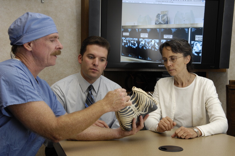
Jane Matsumoto, M.D. (right), co-director of the Anatomic Modeling Lab, with Christopher Moir, M.D. (PHYS ’89) (left), using models to plan the separation of conjoined twins.
Printing 3-D models at Mayo Clinic started in 2008 when surgeons planning the separation of conjoined twins approached the department about producing a replica of the babies’ shared liver, using CT and MRI images as the guide.
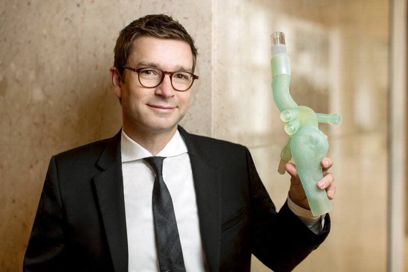
“They had good imaging and illustrations but weren’t entirely clear how the anatomy all worked together,” says Jonathan Morris, M.D. (R-D ’06, R-NEU ’07), Division of Neuroradiology in the Department of Radiology and co-director of the Anatomic Modeling Lab. “We learned that having a model is a critical piece of decision-making, especially in complex surgical cases.”
Jonathan Morris, M.D., co-director of the Anatomic Modeling Lab
In a surgery to separate another set of conjoined twins, the surgical team knew exactly where to cut the shared liver based on 3-D printed models. Mayo’s pediatric spine surgeons saw the models and began requesting them for complex congenital scoliosis cases. Orthopedic oncologists began requesting models for complex tumor resections. Thoracic surgeons followed suit, wanting to see detail including the rib cage, spine, brachial plexus, trachea, tumors, subclavian arteries and veins, and pulmonary vessels. Then it was off to the races, according to Dr. Morris, with a permanent lab established in 2013 on the Saint Marys Campus.
“Mayo is a pioneer in radiology, and we produce great 2D and 3-D imaging,” he says. “But mental gymnastics are required to visualize life-size 3-D objects from 2-D images. Having an actual physical model of the patient’s precise anatomy that you can hold is priceless, especially when working with tumors that change the anatomy. Physicians and surgeons can plan procedures, determine how to place the patient on the operating table and exactly where to cut, and anticipate problems to prevent surprises in the operating room.”
“Having an actual physical model of the patient’s precise anatomy that you can hold is priceless, especially when working with tumors that change the anatomy.”
From the first model in 2008, Mayo’s 3-D printing capabilities have grown significantly. Today a fleet of three industrial 3-D printers runs continuously in the Department of Radiology. A 3-D printed model can take between one hour and three days to print, depending on the size, level of accuracy and materials. Models can be made from plastic or flexible materials in different colors to represent various structures. At present, models can’t simulate the feel of an organ — how the bowel feels different from a kidney, for example — but Mayo is working with vendors to develop even more life-like materials.
Changing the surgical landscape

Thoracic surgeon Shanda Blackmon, M.D.
Having 3-D printed models also has allowed Mayo surgeons to devise less-invasive surgical methods. Shanda Blackmon, M.D. (TS ’16), Division of Thoracic Surgery at Mayo Clinic in Rochester, laparoscopically removed a Pancoast tumor, a rare form of lung cancer, for what is believed to be the first time. The tumor was wrapped around several critical nerves and blood vessels. The patient went home three days later instead of having his chest split open in a traditional operation and requiring a week or more in hospital. [For information about this procedure]
“The orthopedic, vascular and thoracic surgeons all studied the model to plan exactly how we could remove the tumor without a major incision, which wouldn’t have been possible without the model,” says Dr. Blackmon. “3-D printing is changing the landscape of oncologic surgery.”
“3-D printing is changing the landscape of oncologic surgery.”
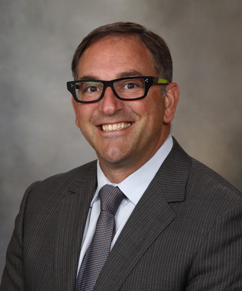
Vascular surgeon Gustavo Oderich, M.D.
Gustavo Oderich, M.D. (S ’04, VASS ’06), Division of Vascular Surgery, pioneered a new type of complex stent to treat aortic aneurysms and placed them in a Mayo Clinic patient — for the first time in the U.S. With a patient-specific 3-D printed model hooked up to a pump system, Dr. Oderich and his team rehearsed every step of the procedure two times, including placing the eight custom sections of stent, before the patient’s surgery. [For information about this procedure]
At Mayo, 3-D printing also is changing the way images are obtained by CT and MRI. “We’re designing imaging protocols specifically geared toward 3-D printing to make sure the original images are tailored for printing when we know we’ll want to create a model,” says Dr. Morris.
The lab began 3-D printing for all Mayo locations at the end of 2016 and has offered a CME course, Collaborative 3-D Printing in Medical Practice, for the last three years. Every week the lab fields calls from institutions inquiring about how to set up a similar lab.
Saving money and time
To date, insurance companies aren’t paying for 3-D models. However, Mayo Clinic sees them as a significant cost-saving tool. An operating room bills at $80 to $150 per minute, and complex surgeries can take hours. The preoperative planning the 3-D printed models allow shaves time off of OR use, making the cost of the model relatively trivial, according to Dr. Morris.
The preoperative planning the 3-D printed models allow shaves time off of OR use, making the cost of the model relatively trivial.
The 3-D models also are invaluable in patient education, helping patients understand the surgical plan along with risks and benefits.
“We live in a 3-D world,” says Jane Matsumoto, M.D. (MED ’80), co-director of the Anatomic Modeling Lab. “Having exact 3-D models of the anatomy to work with means you don’t have to wonder what size something is or what structures are in the way of the surgeon’s path. Our surgeons can hold the model, turn it, rotate it and know what they’re going to do before the patient enters the OR.”
“Our surgeons can hold the model, turn it, rotate it and know what they’re going to do before the patient enters the OR.”