A new breast imaging technique pioneered at Mayo Clinic nearly quadruples detection rates of invasive breast cancers in women with dense breast tissue, according to the results of a major study published this week in the American Journal of Roentgenology.
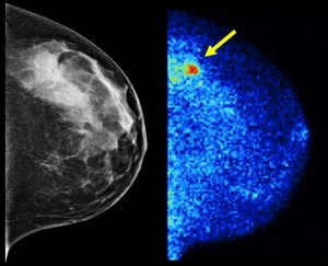
Molecular Breast Imaging (MBI) is a supplemental imaging technology designed to find tumors that would otherwise be obscured by surrounding dense breast tissue on a mammogram. Tumors and dense breast tissue can both appear white on a mammogram, making tumors indistinguishable from background tissue in women with dense breasts. About half of all screening-aged women have dense breast tissue, according to Deborah Rhodes, M.D. (GIM ’97), a Mayo Clinic Breast Clinic physician and the senior author of this study.
MBI increased the detection rate of invasive breast cancers by more than 360 percent when used in addition to regular screening mammography, according to the study. MBI uses small, semiconductor-based gamma cameras to image the breast following injection of a radiotracer that tumors absorb avidly. Unlike conventional breast imaging techniques, such as mammography and ultrasound, MBI exploits the different behavior of tumors relative to background tissue, producing a functional image of the breast that can detect tumors not seen on mammography.
The study, conducted at Mayo Clinic, included 1,585 women with heterogeneously or extremely dense breasts who underwent an MBI exam at the time of their screening mammogram.
- Of these women, 21 were diagnosed with cancer — five through mammography alone (24 percent or 3.2 cancers per 1,000 women) and 19 with mammography plus MBI (91 percent or 12 cancers per 1,000 women).
- Particularly notable was the four-fold increase in detection of invasive cancers (1.9 invasive cancers per 1,000 women with mammography and 8.8 per 1,000 women with mammography plus MBI). Detection rates for noninvasive cancers were not significantly different.
- The risk of incurring an unnecessary biopsy because of a false positive exam increased in this study, from 1 in 100 women with mammography to 4 in 100 women with mammography plus MBI. (By comparison, recent studies have shown that alternative supplemental screening techniques, such as ultrasound and MRI, generate about eight additional unnecessary biopsies per 100 women.)
This information was originally published in a Mayo Clinic press release on January 23, 2015.